Function of the Human Spine: Anatomy 101
If you’re reading this, you’ve likely dealt with back or neck pain, or you want to understand how your spine works so you can help prevent future episodes. Many patients say their previous doctors never really explained what was happening – they had X-rays, MRIs and CT scans without ever understanding the findings. The more you know about your body and how it works, the more active a role you can take in healing.
This article walks through the anatomy and function of the human spine, one structure at a time.
The Vertebral Column
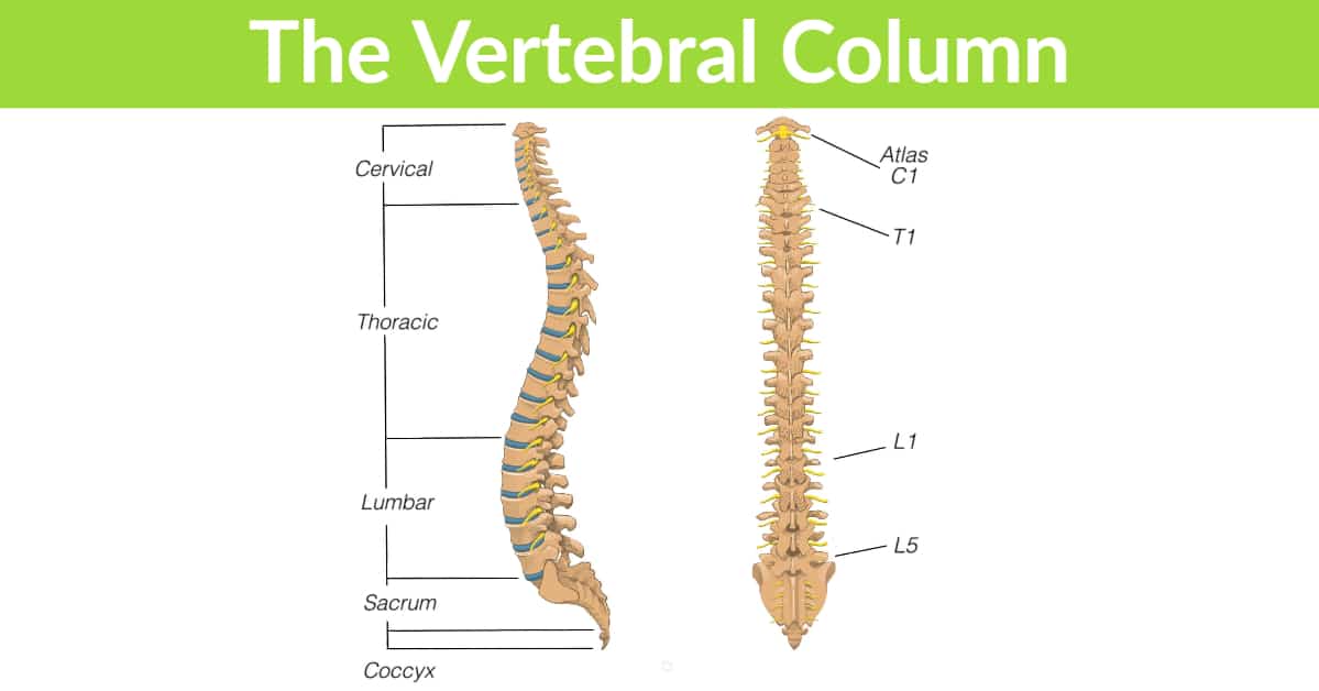
The spine, or vertebral column, is made up of individual bone segments called vertebrae, stacked one on top of another and separated by small, shock-absorbing structures called intervertebral discs. Its job is to support the body’s weight, protect the spinal cord, and enable upright human function in standing, sitting, walking and every other daily activity.
A total of 33 vertebrae form the spine, divided into sections based on the curves they create: the cervical spine (neck), thoracic spine (mid and upper back), lumbar spine (lower back), and the sacrum and coccyx (tailbone). The sacrum and coccyx are unique in that their vertebrae are fused, with no discs between them. The sacrum forms the solid base of the spine, meeting the pelvic bones to form the pelvis – a strong foundation that supports the upper body’s weight and transfers it into the legs.
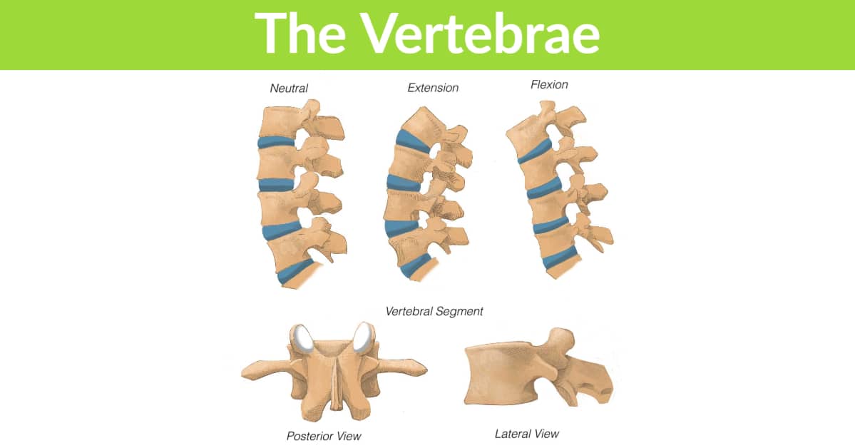
The Vertebrae
Each bony segment of the spine is a vertebra. The front portion, called the body, is the main weight-bearing structure. The back portion is a ring-like structure that forms a hollow canal through which the spinal cord passes. This is part of why spinal alignment and chiropractic care matter – the vertebrae help protect and house the cord and nerves.
The back of each vertebra also includes two facet joints. Like any joint, these have a cartilage surface and are susceptible to wear and tear that can lead to degenerative changes such as arthritis. Vertebrae also attach to ligaments and muscles that connect and anchor them from above and below, with ligaments acting as cable-like structures that hold bones together.
The Spinal Cord and Nerves
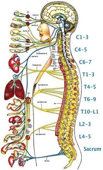
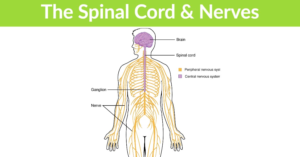
The spinal cord is essentially an extension of the brain, and the nerves that exit it reach out to connect with every major tissue and organ in the body. The brain is the body’s central processing system, coordinating nearly every physiological reaction, from temperature, touch and pain to movement, digestion, breathing and heart rate. The spinal cord is the bundle of nerves that carries these signals to and from the brain – think of it as a protected nerve highway running through the center of the vertebral column.
Together, the spinal cord and brain form the central nervous system (CNS), which is vital to every function in the body. Along the cord’s length, nerves exit through small openings between the vertebrae called intervertebral foramina, specific to each spinal level (for example, between C5 and C6 or L4 and L5). The cervical foramina carry signals to the arms, the lumbar foramina to the legs, and exit points throughout the spine relay signals to and from the body’s organs, including the heart and lungs.
Where the nerves branch off and exit the spine, they form the peripheral nerve roots that make up the peripheral nervous system (PNS). It is at these exit points that many problems arise, because the nerve roots are prone to compression and stretching by nearby structures such as discs, ligaments and bone spurs. Nerves under sustained pressure can cause a range of issues, which is why maintaining (or restoring) spine health is so important.
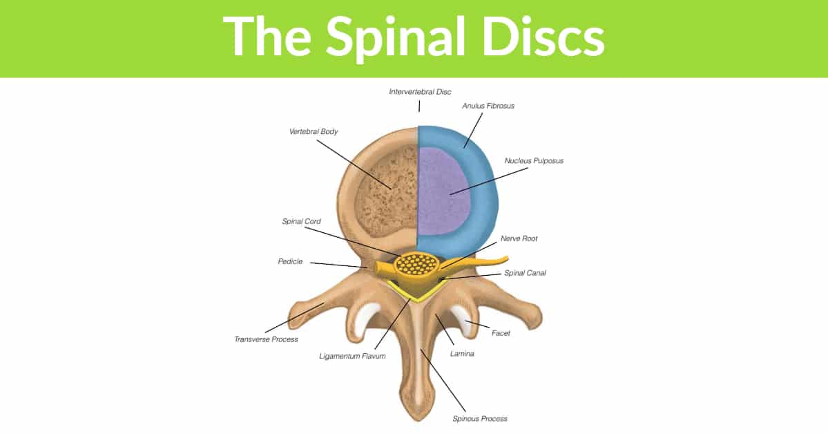
The Spinal Discs
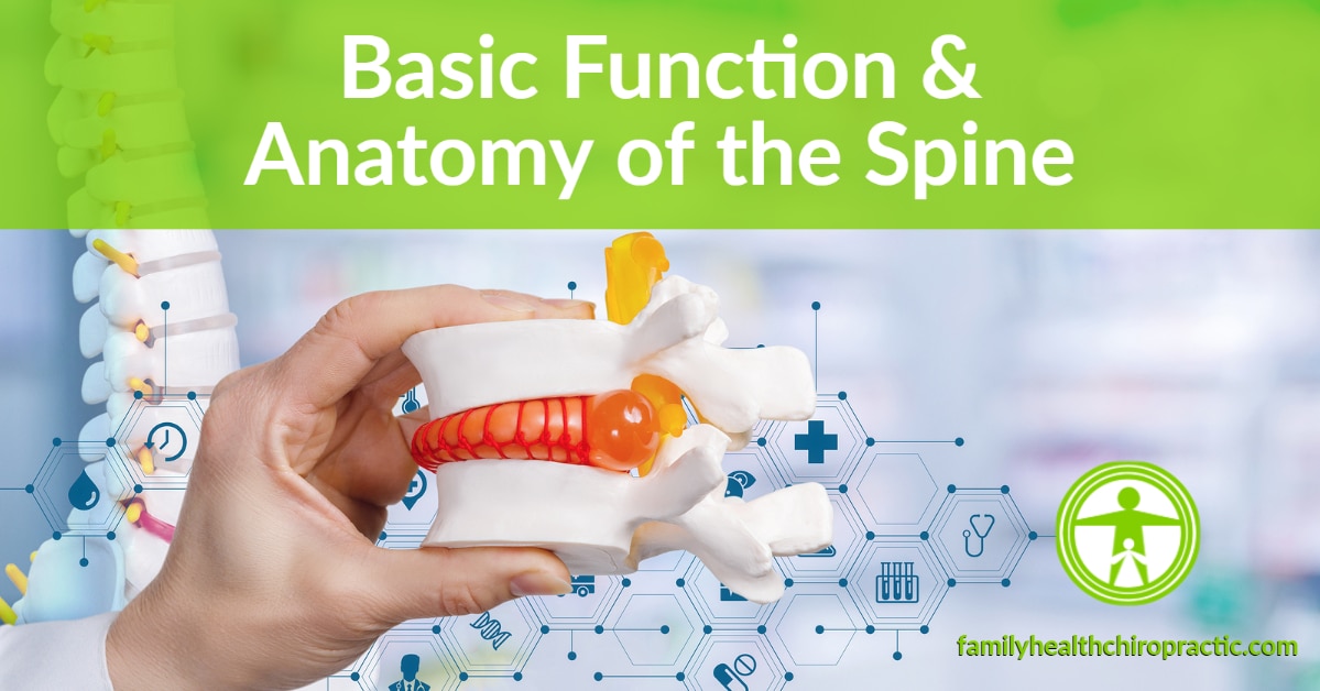
Discs probably get more attention than any other spinal structure – usually in their damaged state, as bulging, ruptured or herniated discs. A disc is an elastic structure between two vertebrae that separates them and acts as a shock absorber. A common analogy is a tough jelly donut: a gooey center (the nucleus pulposus) surrounded by tough, fibrous collagen rings (the annulus). The nucleus is a blend of fibers, sugars and water that gives it a sponge-like, hydrodynamic quality.
To maintain that water-absorbing function, the gel needs nutrients. In early life, blood supply through the vertebrae above and below feeds the disc. But as we mature, the growth plates close and cut off that direct blood supply. From then on, the disc receives nutrients through a pump-like process of alternating compression and relaxation, which draws nutrients in and out of the nucleus from the surrounding tissue.
When the inner gel is well hydrated, it works like a properly inflated tire – internal pressure pushes the annulus fibers outward, keeping them elongated and able to absorb shock. Now imagine what happens when a disc doesn’t get that essential compression-and-relaxation cycle. Sitting all day compresses discs constantly with little chance to decompress. Add slouching plus repeated bending and twisting, and you have an under-pressurized disc that can overstretch and tear – the world of bulging and herniated discs.
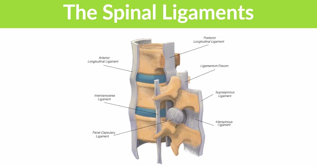
The Spinal Ligaments
Ligaments are cable-like structures that connect bone to bone. They are slightly elastic and can stretch a little, but if overstretched they lose their stiffness and fail. They also have a sensory role (proprioception): their nerve endings signal the brain when stretched, prompting supporting muscles to contract and stabilize the joint. They are critical to the structural integrity of every joint in the body.
Ligaments are especially important in the spine because of its long, vertical length and many movable segments. Picture building a skyscraper without support beams – it would collapse. Each spinal ligament helps keep motion in check; if even one fails, it can throw off the balance of forces and stress the surrounding discs, joints and ligaments. Because the spine moves in many directions, it has ligaments attaching at various levels and angles – for example, the posterior longitudinal ligament running down the back of the vertebrae helps limit forward bending, while the anterior longitudinal ligament on the front helps limit backward bending. The key point: prolonged poor posture and movement can overstretch ligaments until they fail, at which point muscles must step in to provide support.
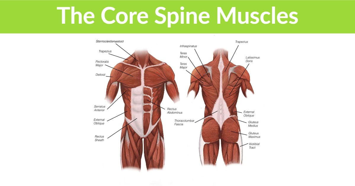
The Core Spinal Muscles
The core is often misunderstood as just the abdominal muscles, but it is much more than that. It includes the chest, back and abdomen along with the spine, rib cage, and the pelvic and shoulder girdles – essentially the link between the upper and lower body and all the muscles spanning that distance.
The core muscles work together as an integrated system, much like musicians in an orchestra. They keep us upright, resist compression, support body weight and provide the framework for posture during every movement, from pushing and pulling to squatting and bending. One of their main jobs is to keep the spine stable so the powerful hip and leg muscles can transfer force into the shoulders and arms, and vice versa. Picture a tennis serve: power begins by bending the hips and knees, then recoils upward, transferring force through the core into the shoulders and arms and finishing with a snap at the wrist. Even walking relies on the core to complete this energy transfer. How much each muscle contracts and stiffens its joints determines the overall stability of the spine.
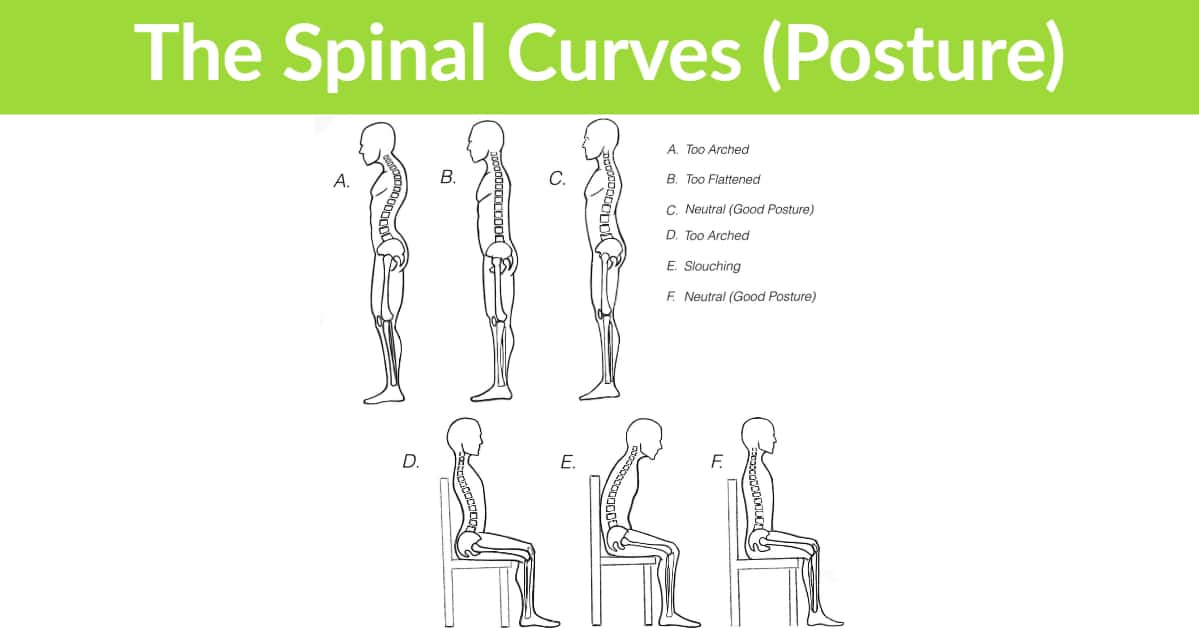
The Spine’s Curves (Posture)
Finally, the spine isn’t actually vertical – it is made up of curves. Viewed from the side in an upright position, these curves define human posture. The spine has three main curves corresponding to the cervical, thoracic and lumbar regions. They develop in infancy, shaped by muscle contractions and the wedge-like shape of the discs. In a healthy spine, the curves balance one another around the body’s center of gravity.
These curves are essential to upright function. Their arch-like design gives the spine strength to resist gravity’s compressive forces with minimal energy – much like a suspension bridge built to support enormous weight. Unfortunately, prolonged poor posture and repeated bad movements can change these curves, flattening or exaggerating them. The low back and neck are especially prone to flattening with age as the structures wear down from poor positioning.
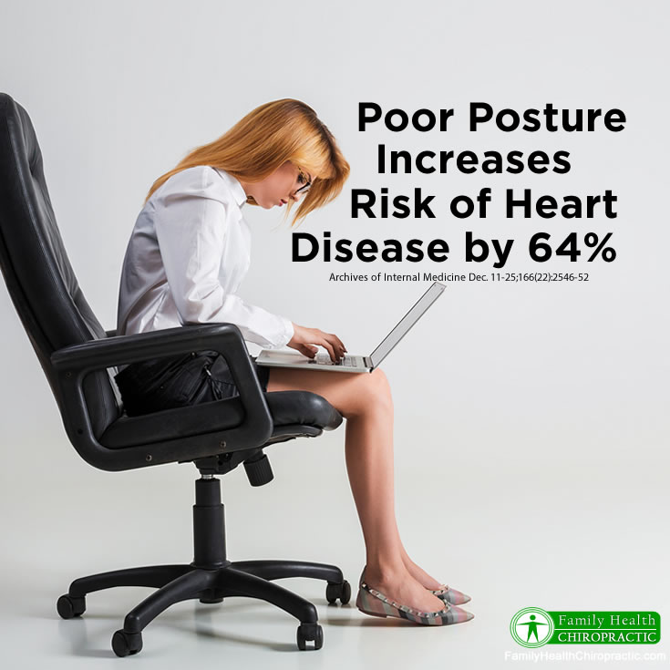
Consider how many years someone might spend sitting slouched, texting with the head down, or bending poorly. In these situations the ligaments, discs and joints are overstressed and can fail, muscles stop doing their jobs, and the spine can no longer stay erect or absorb load well. The whole core system is compromised, and efficient movement declines – a negative feedback loop that wears the body down. The good news is that the reverse is also true: the right exercises, movement patterns and postures can rebuild the core and take stress off the spine.
READY TO FEEL BETTER?
Book Your Appointment Today
Same-week appointments are often available. Call 512-347-8881 or request a time online.
Ready to feel better?
If you are new to our office, learn what to expect on your first visit, or see our full range of chiropractic services.